Sorry for the delay
 Margaret
Things have been moderately insane recently. But I promised to explain the previous radiographs so here goes.

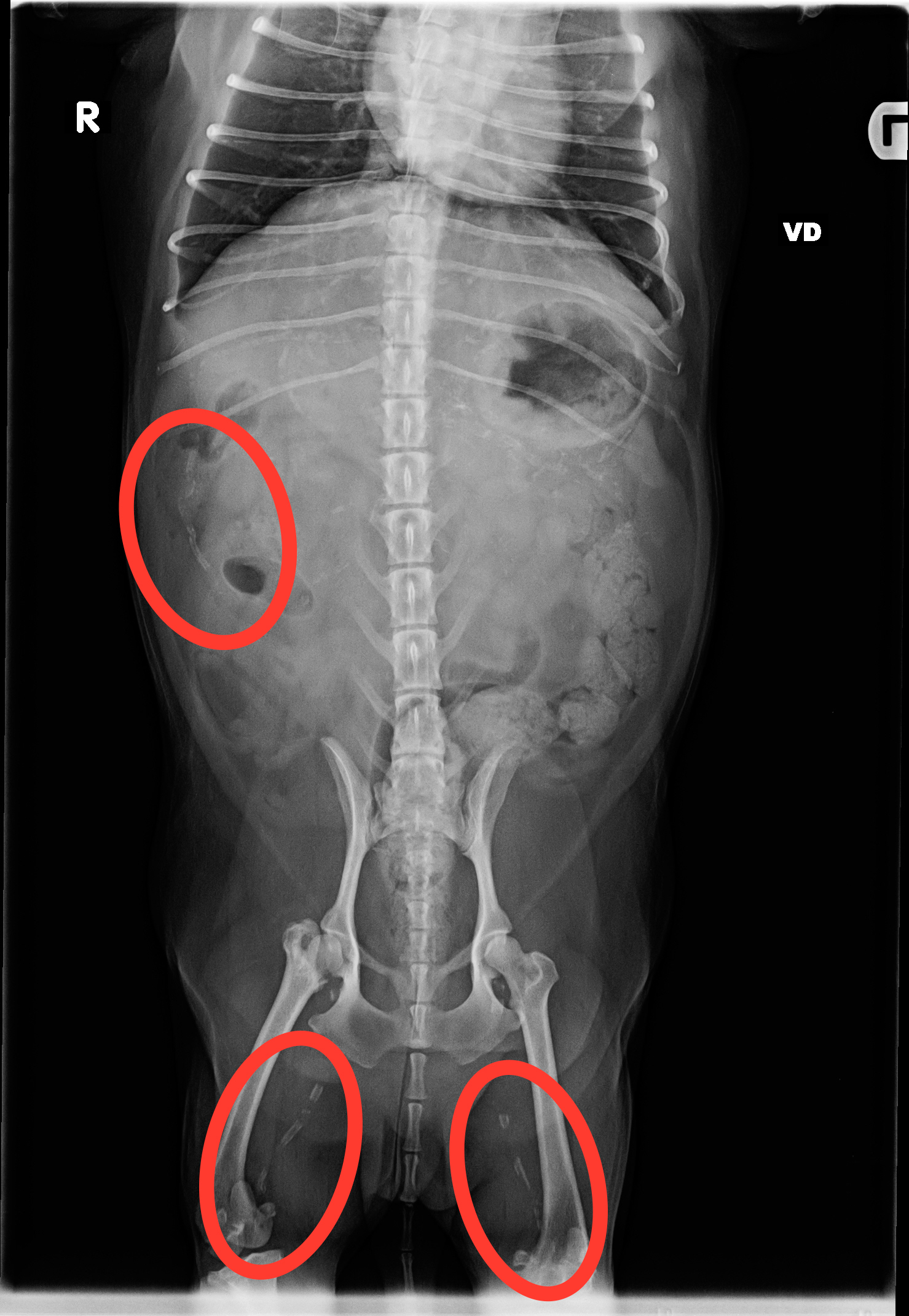
So this image is a ventrodorsal (he’s lying on his back) image of Andy’s abdomen. Andy’s head is towards the top, his butt is towards the bottom of the images. The circles are pointing out Andy’s calcified right renal artery (the little squiggly white line in the center of the single circle camera left), and Andy’s two partially calcified femoral arteries (the little white linear blotches in the center of the circles near the bottom of the image).

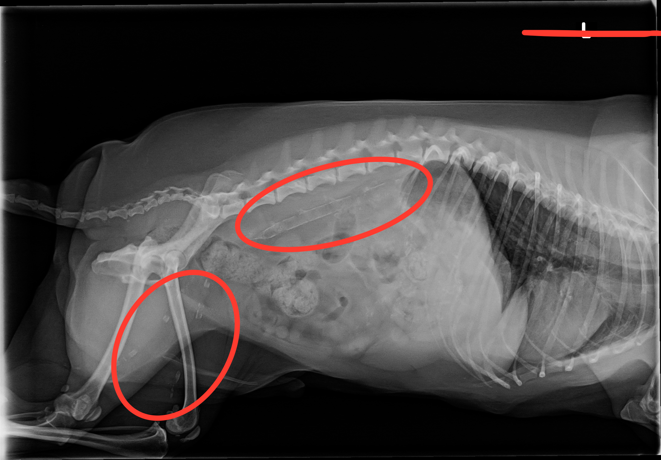
This is a left lateral abdominal radiograph. Andy is lying on his left side and his head is towards your right. The big oval circle near the top is pointing out the calcified portion of Andy’s terminal aorta (complete with calcifications of the major arteries that come off the aorta) and the other circle lower down is pointing out the calcified femoral arteries again.
The big red line at the upper right of the image is an oops with the highlighter feature of the image software that I can’t figure out (nor do I want to try) how to get rid of. Hey, I deal with carbon based life forms, not silicone ones.
Seeing these images come up after I’d taken the radiographs was one of those professional “Oh holy SHIT I’ve never seen THAT before!” moments that happen quite rarely. And since I managed to wig out both a board certified internal medicine specialist and a board certified radiology specialist with these images, I’d bet that I’m never going to see another one of those again. No, I don’t know why those blood vessels are suddenly becoming calcified. I’m currently working with both a wigged out board certified neurology specialist and a wigged out board certified surgeon to try and get an answer.
Carbon based life forms are such a glorious mystery.


